Figure 1.

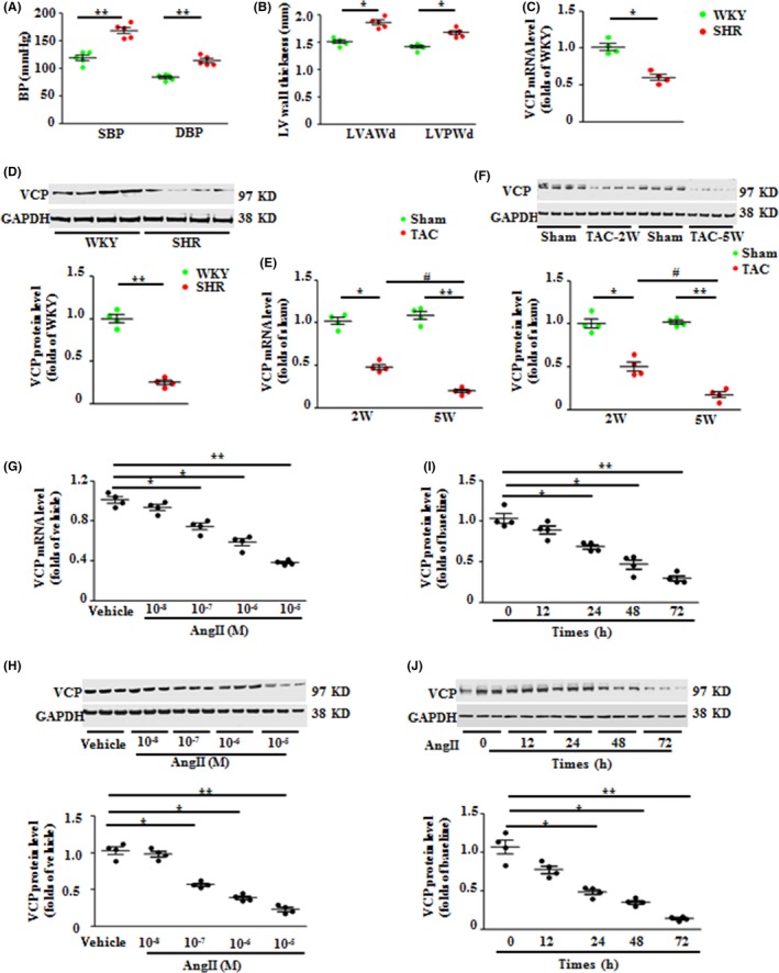
Valosin‐containing protein (VCP) expression is downregulated in hypertrophic hearts upon pressure overload. (A) The systolic and diastolic blood pressure (SBP and DBP) of Wistar Kyoto (WKY) rats and spontaneously hypertensive rats (SHR). (B) The left ventricle (LV) anterior or posterior wall thickness of end‐diastolic phase (LVAWd) and (LVPWd) of rat hearts. The mRNA (C) and protein levels (D) of VCP in the rat LV tissues. n = 4–5/group. *P < 0.05, **P < 0.01 vs. WKY. (E–F) The mRNA (E) and protein levels (F) of VCP in the LV tissues of wild‐type (WT) mice under transverse aortic constriction (TAC) for 2 weeks and 5 weeks. *P < 0.05, **P < 0.01 vs. sham; # P < 0.01 vs. 2‐weeks TAC mice. n = 4/group. (G–J) The mRNA and protein levels of VCP in cultured neonatal rat cardiomyocytes (NRCMs), under the treatment of angiotensin II (AngII) with different doses or with vehicles (Veh) for 48 h, or at different time courses at the dose of 10−6 m. *P < 0.05, **P < 0.01 vs. vehicle control. Representative blots showed the examples of three of four experiments from each group. n = 4 sets independent experiments/treatment for quantitation, each experiment was performed in triplicate and averaged. GAPDH is a loading control for total protein. Data are shown as mean ± SEM, one‐way ANOVA was used.
