Figure 5.

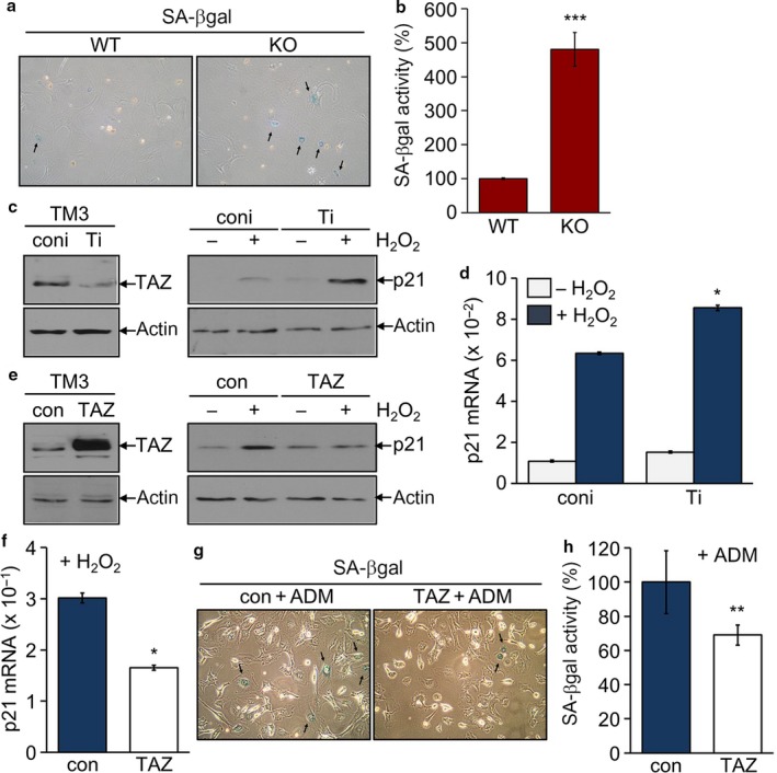
Suppression of p21 expression and senescence by TAZ. (a, b) Primary testicular cells isolated WT and TAZ KO mice (12 months old, n = 6 each group) were maintained for three passages. Cells were subjected to a SA‐βgal staining (a) and β‐galactosidase activity assay (b). Data are mean ± SD of six experiments. ***P < 0.0005. (c–h) Stable cell lines were established in mouse TM3 cells: knock‐down control (coni), TAZ knock‐down (Ti), control (con), and TAZ stable cells (TAZ), and treated with 100 μM H2O2 for 4 h. Immunoblotting analysis of TAZ, p21, and actin (c, e). Quantitative analysis of p21 mRNA level (d, f). *P < 0.05. Stable cells, con and TAZ, were incubated with 50 nM ADM for 3 days and assayed with a SA‐βgal staining (g) and β‐galactosidase activity assay (h). **P < 0.005.
