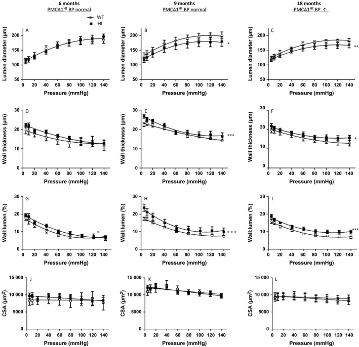
Figure 3.

Remodelling of small mesenteric arteries occurs with age in PMCA1Ht mice and before a detectable increase in BP. (A) The internal lumen diameter does not significantly differ between passive mesenteric arteries from WT and PMCA1Ht mice at 6 months of age but is significantly smaller in PMCA1Ht mice at 9 and 18 months (B & C). (D) Wall thickness is similar at 6 months of age and significantly increases with age in PMCA1Ht arteries (E & F). (G, H. and I) The vessel wall thickness to lumen ratio (W:L) is significantly increased in arteries from PMCA1Ht mice aged 6, 9 and 18 months old. (J, K and L) The cross‐sectional area (CSA) of the vessel wall is not significantly different between arteries taken from WT and PMCA1Ht mice at any age tested. Extra sum of squares F‐test analysis was performed. All data were plotted as mean value ± SEM. n = 4–8 *P < 0.05, **P < 0.01, ***P < 0.001.
