
Figure 5.
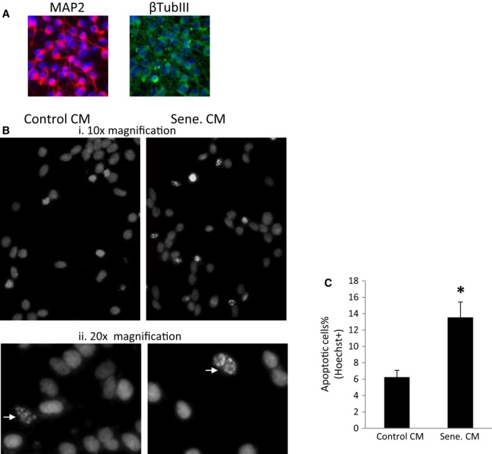
Conditioned medium of senescent HFAs induces neurotoxicity. (A). differentiated LUHMES cells at day 5 were stained for MAP2 and the neurofilament tubulin III protein. B, HFAs were treated with the control vehicle or meth to induce senescence. LUHMES cells were differentiated into human dopaminergic neurons for 2 days. At day 3, media were changed to the HFA conditioned media. At day 5, neurons were stained with Hoechst dye. Apoptotic neurons were detected as fragmented nuclei staining, indicated by arrows. The apoptotic neurons were imaged and quantitated (C). * denotes P < 0.05. n = 4.