
Figure 6.
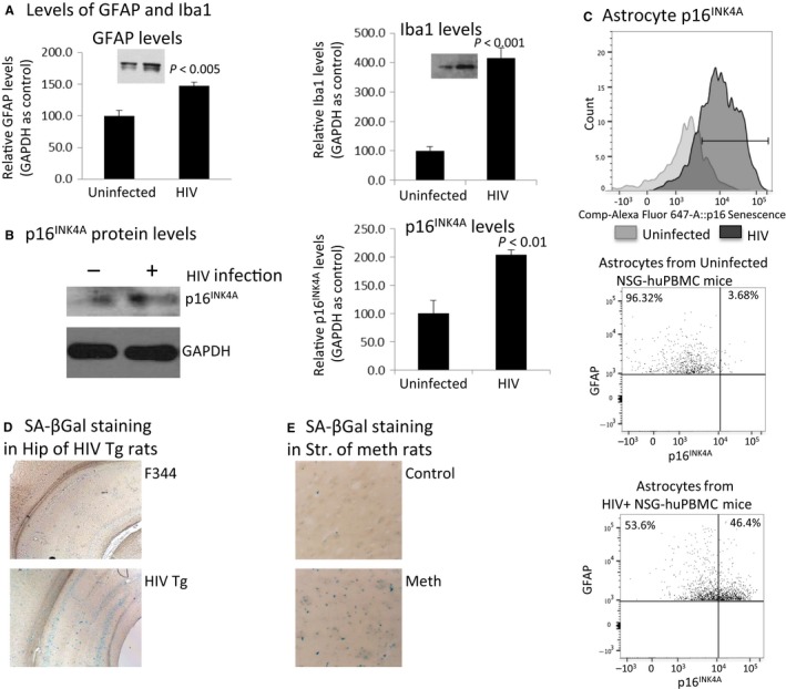
In vivo detection of astrocyte senescence mediated by HIV or meth in three small animal models. A). Brains from HIV+ NSG‐huPBMC mice (n = 8) were dissected and brain lysates were extracted with 1xRIPA buffer and analyzed for the levels of GFAP (A) and Iba1 (B), and p16INK 4A (C) by Western blotting. In (D), astrocytes were isolated by Percoll gradient after enzymatic digestion of brain tissue. The astrocytes were immunostained with anti‐GFAP and anti‐p16INK 4A and further labeled with appropriate secondary antibodies conjugated with Alexa‐594 and Alexa‐488, respectively. Astrocytes were then analyzed by flow cytometry. E, brains (n = 8) were harvested from adult male HIV‐Tg and non‐Tg rats and stained for SA‐βGal. Images were representative staining in the cortex. F) Adult Sprague Dawley rats were treated with a meth regimen (10 mg/kb, i.p., every 2 h × 4). At day 7, brains were fixed and stained for SA‐βGal. Images were representative staining of SA‐βGal in the striatum.