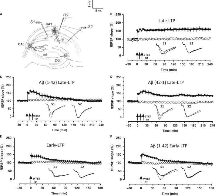
Figure 1.

Aβ 1–42 impairs late‐LTP but not early‐LTP. (A) Schematic representation of the positioning of electrodes in the CA1 region of a transverse hippocampal slice. Recording electrode (rec) positioned in CA1 apical dendrites was flanked by two stimulating electrodes S1 and S2 in stratum radiatum (sr) to stimulate two independent Schaeffer collateral (sc) synaptic inputs to the same neuronal population. (B) Application of strong tetanization (STET) in S1 (filled circles) resulted in late‐LTP. The control potentials in S2 (open circles) were relatively stable (n = 6). (C) Hippocampal slices pretreated with Aβ 1–42 (Aβ, 200 nm) for 2 h during the incubation period failed to show late‐LTP after STET in S1 (filled circles) (n = 8). (D) Aβ 42–1 (200 nm)‐treated slices expressed late‐LTP after the application of STET (n = 9). (E–F) Induction of early‐LTP in S1 (filled circles) using a weak tetanization (WTET) protocol in both the control slice (n = 6) and Aβ‐treated slices (n = 7) resulted in early‐LTP. Control potentials from S2 remained stable during the recorded period (open circles) in all the cases. Analog traces represent typical field EPSPs of inputs S1 and S2 15 min before (solid line), 30 min after (dotted line) tetanization and at the end of the recording (dashed line). Solid arrow indicates the time point of STET or WTET of the corresponding synaptic input. All data are plotted as mean ± SEM. Error bars indicate SEM. Calibration bar for all analog sweeps: 3 mV per 5 ms.
