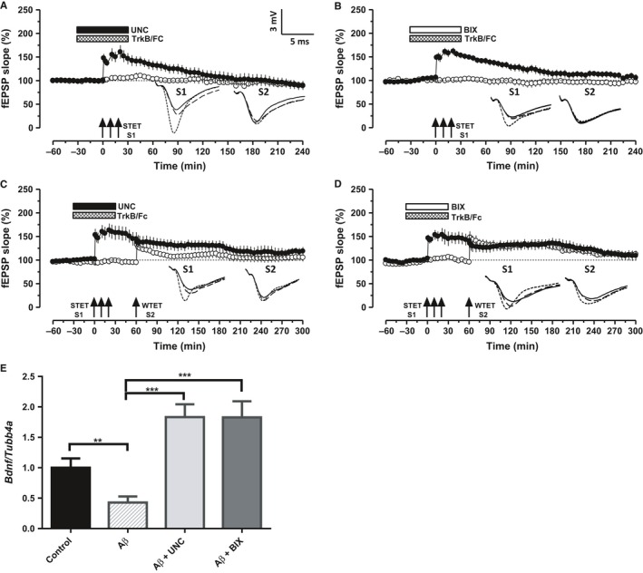
Figure 5.

BDNF mediates the restoration of Aβ induced deficits in LTP and STC during the inhibition of G9a/GLP complex. A–B. Application of STET in the presence of BDNF chelator TrkB/Fc (1 μg mL−1) and either of the G9a/GLP inhibitors UNC (A, n = 7) or BIX (B, n = 7) failed to express a long‐lasting LTP (filled circles) in the Aβ‐treated slices. Control potentials (open circles) were stable throughout the recording. C–D. Inhibition of G9a/GLP complex failed to rescue Aβ‐induced deficits in STC when BDNF is chelated using TrkB/Fc. STET in S1 (filled circles) in the presence of UNC (C, n = 7) or BIX (D, n = 6) along with TrkB/Fc resulted in early‐LTP. Induction of early‐LTP in S2 60 min after the application of WTET in S1 also resulted in early‐LTP (open circles), hence no expression of STC. (E) Bdnf mRNA is downregulated in the Aβ‐treated hippocampal slices (P < 0.01). Increased relative expression of Bdnf mRNA in CA1 region of Aβ‐treated hippocampal slices after UNC/BIX application for 1 h (one‐way ANOVA, F = 12.71; P < 0.001). The values of the individual groups were calculated in relation to the control group. Each bar represents mean ± SEM (n = 6). Asterisk indicate significant difference (Bonferroni post hoc test, ***P < 0.001). Symbols and analog traces as in Fig. 1.
