
Figure 8.
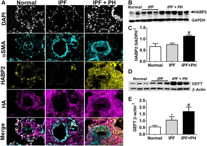
Increased vascular HABP2 is observed in patients with IPF and IPF + PH. Lung sections from a pulmonary vessel showing triple immunofluroescent staining for αSMA (turquoise signals), HABP2 (yellow signals) and HA (violet/purple signals) and counterstained with DAPI (white/grey signals) (A); immunoblots for HABP2 and GADPH (B); densitometry for HABP2 normalized to GADPH (C); immunoblots for GEFT and β‐actin (D); and densitometry for GEFT normalized to β‐actin (E) from normal lung tissue and from lung explants from patients with IPF and IPF + PH. The scale bar represents 50 μm. * P ≤ 0.05, significant difference between normal and IPF + PH values.