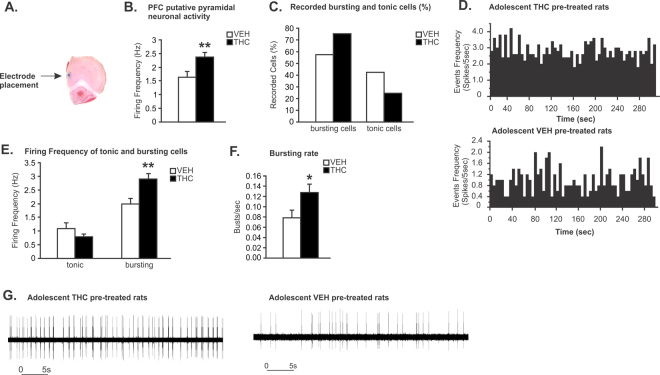
Figure 2.
Long-term effects of chronic THC exposure during adolescence on spontaneous mPFC putative pyramidal neuron activity. (A) Microphotograph of a representative mPFC neuronal recording placement. (B) Adolescent THC pretreated rats displayed increasing spontaneous PFC putative pyramidal neuronal firing frequency. (C) Greater proportion of bursting neurons was observed in adolescent THC exposed rats when compared to VEH controls (75.41% vs. 57.58%). (D) Representative rastergrams showing spontaneous activity of putative PFC pyramidal neurons in THC (top) vs. VEH pretreated rats (bottom). (E) Firing frequencies of bursting cells, not tonic cells, were significantly higher in adolescent THC exposed rats when compared to VEH controls. (F) In the bursting cells population, the bursting rate of putative pyramidal neurons of adolescent THC-exposed rats was significantly higher than in VEH controls. (G) Representative examples showing bursting activity of putative PFC pyramidal neurons in THC (left) vs. VEH pretreated rats (right). Two-tailed t-tests; **indicated p < 0.01; *indicated p < 0.05. Error bars represent the standard error of the means (SEMs).

