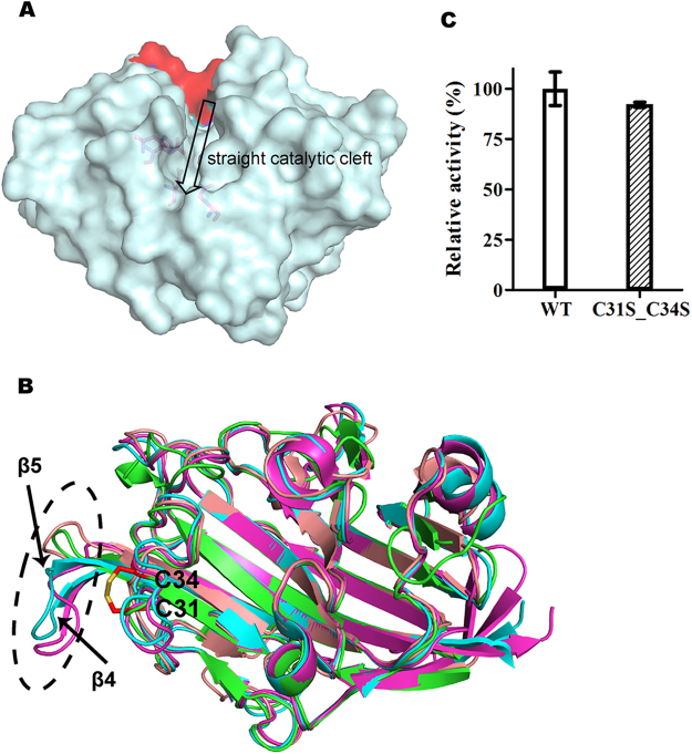
Figure 2.
(a) The surface of ULam111 displaying a straight topology. The additional loop of ZgLamA is red. (b) Superposition of the structures of ULam111 (green), BglF from Nocardiopsis sp. strain F96 (salmon), TmLam (cyan), and PfLamA from hyperthermophile Pyrococcus furiosus (magenta). The disulfide linkage of Cys31-Cys34 is shown as red sticks. The black dashed cycles showed different structures between ULam111 and other GH16 family enzymes. (c) Relative activity of ULam111 mutant toward laminarin. The activity of wild-type ULam111 is represented as 100 and the error bars are standard deviations (n = 3).

