Figure 1.
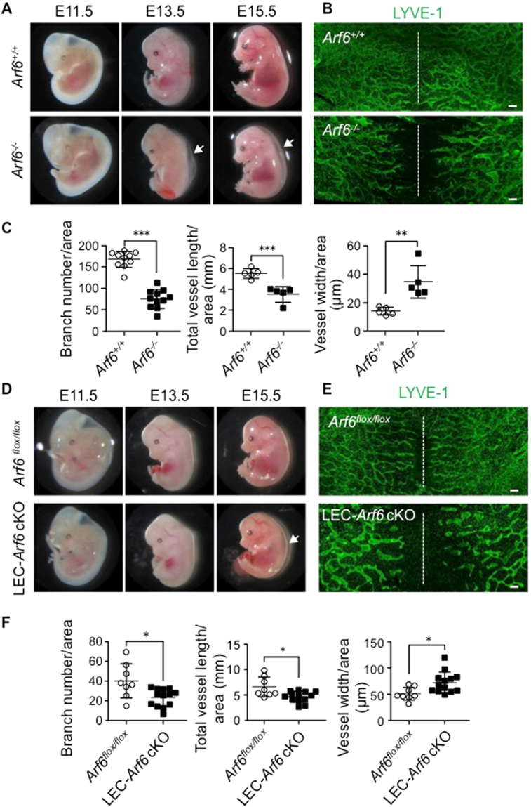
Arf6 −/− and LEC-Arf6 cKO mice induce dorsal skin edema and abnormal lymphatic vascular network. (A,D) Appearance of Arf6 −/− and LEC-Arf6 cKO embryos in comparison with that of control Arf6 +/+ and Arf6 flox/flox embryos, respectively. Note that the edema (white arrows) on the back of E13.5 and 15.5 Arf6 −/− embryos and of E15.5 LEC-Arf6 cKO was induced. (B,E) Aberration of dorsal subcutaneous lymphatic vascular network in E15.5 Arf6 −/− and LEC-Arf6 cKO embryos. Lymphatic vessels were immunostained for LYVE-1 (green). White dashed lines indicate the dorsal midline of the embryo. (C,F) Immunostained images of lymphatic vessels shown in (A) were quantified for branch number of lymphatic vessels/area (left panel), lymphatic vessel length/area (middle panel), and width of lymphatic vessels/area (right panel) in control Arf6 +/+ and Arf6 −/− embryos (C) and control Arf6 flox/flox and LEC-Arf6 cKO embryos (F). Area of 2250 × 1700 μm on both sides of the midline was measured. Each point represents individual value: n = 10 for both embryos in the left panel and n = 5 for both embryos in the middle and right panels of (C), and n = 8 for Arf6 flox/flox embryos and n = 13 for LEC-Arf6 cKO embryos in (F). Statistical significance was assessed using student’s t-test. *P < 0.05, **P < 0.01, ***P < 0.005. Scale bar, 200 μm.

