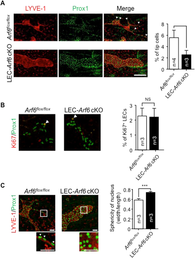
Figure 3.
Arf6 regulates lymphatic vessel sprouting and tip cell morphology. (A) Representative images of sprouting lymphatic vessels in E15.5 control Arf6 flox/flox (n = 4) and LEC-Arf6 cKO (n = 3) (left panels). Transverse jugular sections were co-immunostained for LYVE-1 (red) and Prox1 (green). Arrowheads indicate sprouts of lymphatic vessels. Immunostained images shown in the left panels were quantified for the number of Prox1+ lymphatic tip cells in the distal migration front area of lymphatic vessels (right panel). Scale bar, 200 μm. (B) Representative images of proliferating mLECs in the subcutaneous lymphatic vessel of E15.5 control Arf6 flox/flox (n = 5) and LEC-Arf6 cKO (n = 5) embryos co-immunostained for Prox1 (green) and Ki67 (red) (left panels). Arrowheads represent Prox1+/Ki67+ proliferating mLECs (left panel). Percentages of Prox1+/Ki67+ proliferative mLECs of total Prox1+ mLECs in the distal migrating front area of lymphatic vessels were quantified (right panel). (C) Representative images of subcutaneous lymphatic vessels in E15.5 control Arf6 flox/flox (n = 3) and LEC-Arf6-cKO embryos (n = 3) co-immunostained for LYVE-1 (red) and Prox1 (green). Lower panels are magnified images of the square area in the upper panels. Arrowheads in the magnified images indicate the oval nucleus. Immunostained images were quantified for sphericity of nucleus (width/length) (right panel). Statistical significance was assessed using student’s t-test. NS, not significant, *P < 0.05, ***P < 0.005. Scale bar, 200 μm (A,B) and 25 μm (C).

