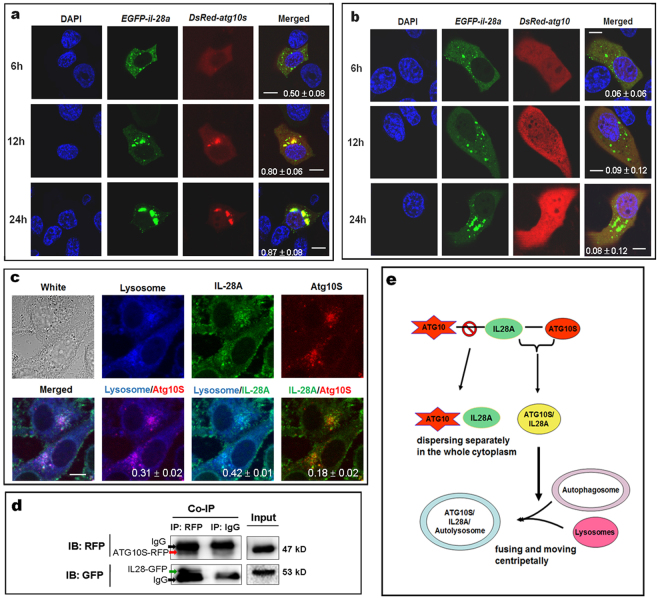
Figure 6.
ATG10S combines to IL28A and fuses to autolysomes but ATG10 not. (a) Co-transfection of egfp-fused il28a expression plasmid and DsRED-fused atg10s plasmid shows that ATG10S had co-localized with IL28A, and the conjunct particles (yellow particles) grew and accumulated at perinuclear region in HepG2 cells in a time-dependent form. (b) Co-transfection of egfp-fused il28a plasmid and DsRED-fused atg10 plasmid did not show IL28A-ATG10 conjugate in the whole experimental process. Scale bars = 10 μm (in a and b) (c) Using lysosome tracker dye, triple colocalization of lysosomes with IL28A and ATG10S was observed in HepG2 cells; two-by-two-merged figures show positive combined particles of lysosome-IL28A, lysosome-ATG10S, and IL28A-ATG10S, respectively. Scale bars = 8 μm (c). The values of Pearson coefficient and the standard deviation are put at the bottom of the merged figures (in a–c). (d) Co-IP test shows the interaction between IL28A and ATG10S using the egfp-fused il28a plasmid and DsRED-fused atg10s plasmid co-transfection in HepG2 cells. Immunoprecipitation with anti-RFP antibody showed combination of ATG10S with IL28A. A red arrow points to the fusion protein of ATG10S-RFP (about 47 kD), and a green arrow points to IL28 fused to GFP (about 53 kD). Black arrows indicate IgG protein (about 50 kD), which is an accompaniment of Immunoprecipitation. Full-length or original blots/gels are presented in Supplementary Figure S11d. (e) A model for IL28A interaction with two ATG10 proteins.

