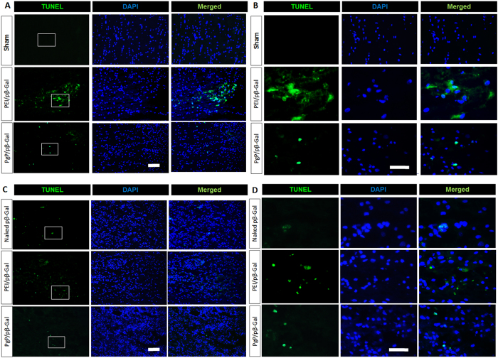
Figure 5.
Cytotoxicity of PgP/pβ-Gal polyplexes at N/P ratio of 30/1 in normal rat spinal cord. At 2 and 7 days post-injection, rats were sacrificed and spinal cords explanted, embedded, sectioned, and toxicity was evaluated by TUNEL staining. Apoptotic cells were stained in green with blue DAPI nuclear counterstaining. (A and B) show representative images of TUNEL+ cells in spinal cord at 2 days post-injection (Top: sham control, Middle: bPEI/pβ-Gal polyplexes at N/P ratio of 5/1, Bottom: PgP/pβ-Gal polyplexes at N/P ratio of 30/1). (C and D) show representative images of TUNEL+ cells in spinal cord at 7 days post-injection (Top: naked pDNA, Middle: bPEI/pβ-Gal polyplexes at N/P ratio of 5/1, Bottom: PgP/pβ-Gal polyplexes at N/P ratio of 30/1), (A and C) Original magnification 200X (scale bar indicates 100 µm), (B and D) Enlarged images of highlighted interest region, original magnification 400X (scale bar indicates 50 µm).

