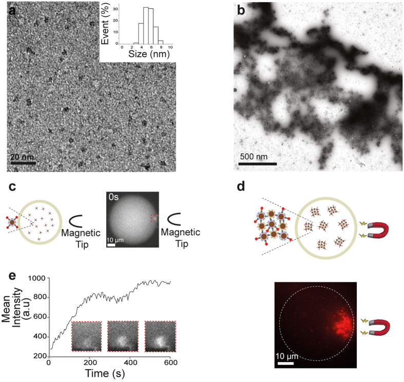
Figure 3.
Characterisation and magnetic manipulation of biomineralised ferritins. (a,b) TEM images of mineralized FRB-ferritins and ferritin clusters (negative staining). Monodisperse iron oxide nanoparticles are shown in (a). The addition of Rapamycin and FKBP-ferritins into the FRB-ferritin solution causes the assembly of proteins nanocages to form micrometer-sized structures (b). (c,d) Fluorescence images of mineralized FRB-ferritins and ferritin clusters. The nanocages were labelled with FKBP-mCherry for fluorescence visualization within droplets of water dispersed within mineral oil (see schematic). Magnetic forces were induced by (c) a magnetic tip for focusing the gradient of forces, or (d) a permanent magnet for a long-range attraction of the clusters. (e) Mean fluorescence intensity as a function of time at the vicinity of the magnetic tip showing the local recruitment of single ferritins.

