Figure 4.
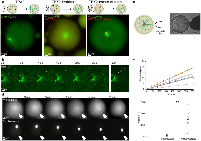
Nucleation and spatiotemporal manipulation of microtubules assemblies upon magnetic actuation. (a) Confocal observation of microtubule polymerization induced by FKBP-TPX2 in a droplet of Xenopus egg extract. Microtubules and ferritins are labelled with fluorescein-labelled tubulin and mCherry, respectively. From the left to right, microtubule structures triggered with FKBP-TPX2, mCherry/TPX2-ferritins, and ferritin-TPX2 clusters. (b) Time-lapse of microtubule growth polymerizing from an aster triggered by ferritin-clusters. EB1-GFP is used as fluorescent reporter of the plus-end microtubule growing extremity. c, Schematic of the magnetic control experiment (left). Bright-field observation of the magnetic tip positioned next to an egg extract droplet (right). (d) Representative time-points of the dynamic of an aster centre upon magnetic field actuation. (e) Example of the spatiotemporal dynamics of aster centres toward the gradient of magnetic field. Plot of the length of the trajectory of the aster as function of time when submitted to magnetic forces. (f) Mean velocity of the asters attracted toward the magnet in absence and in presence of a microtubule-depolymerizing drug (nocodazole).

