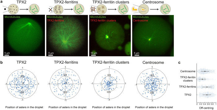
Figure 5.
Ferritin Scaffolding mimics the positioning of Microtubule Organizing Centres of cells. (a) Representative observations of microtubule aster organization triggered by FKBP-TPX2, TPX2-ferritin, TXP2 ferritin-clusters, and centrosomes. Microtubules and ferritins are labelled with fluorescein-labelled tubulin and mCherry, respectively. (b) Geometrical positioning of asters nucleated by FKBP-TPX2, TPX2-ferritin, TXP2 ferritin-clusters, and centrosomes. 0 and ±1 indicate the centre and the droplet boundary, respectively. (c) Mean and standard deviation of the off-centring of the aster positions triggered by FKBP-TPX2, TPX2-ferritin, TXP2 ferritin-clusters, and centrosomes.

