
Figure 2.
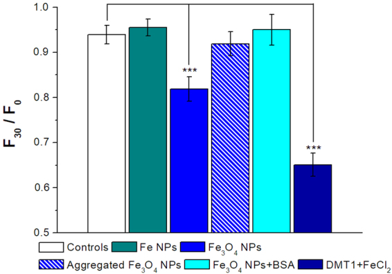
Iron oxide NPs induce intracellular iron increase. Histogram of the means of the F30/F0 values calculated as the fluorescence values at time 30 min normalized for the fluorescence values at time 0. Fe3O4 NPs (blue column) caused a statistically significant quenching of Calcein compared to the controls, i.e., Calcein injected oocytes exposed to external control solution at pH 7.6 (white column). Conversely, Fe NPs (green column), aggregated Fe3O4 NPs (blue barred column) and BSA modified Fe3O4 NPs (cyan column) did not induce quenching; rDMT1 transfected oocytes exposed to iron chloride were used as positive controls (dark blue column). Bars represent ± SEM; from 10 to 40 oocytes for each column deriving from 2 to 8 batches were used. Statistical analysis was performed with One-way ANOVA and orthogonal comparisons with Holm-Bonferroni post hoc test (***p < 0.005).