Figure 3.
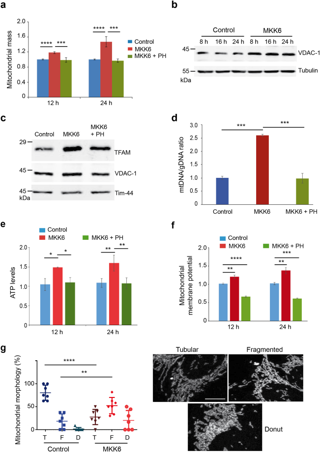
MKK6 expression affects mitochondria function. U2OS cells expressing a Tet-regulated construct were either mock treated (control) or treated with tetracycline for the indicated times to induce the expression of constitutively active MKK6. (a) Mitochondrial mass in control and MKK6 expressing cells was measured using MitoTracker Deep Red at the indicated times, and is represented as fold change versus the control. (b) Total lysates from control and MKK6 expressing cells were analysed by immunoblotting. (c) Mitochondria were extracted from control and MKK6 expressing cells in the presence or absence of the p38α inhibitor PH797804 (PH) for 24 h. Expression of the indicated mitochondrial proteins was analysed by immunoblotting. The uncropped immunoblots are presented in Supplementary Fig. S8. (d) The ratio of mitochondrial DNA (mtDNA) versus genomic DNA (gDNA) was analysed in control and MKK6 expressing cells in the presence or absence of PH for 24 h. (e) ATP levels were analysed at the indicated times after MKK6 induction in the presence or absence PH and were normalized towards the cell number. Values were standardised towards control cells. (f) Mitochondrial membrane potential in control and MKK6-expressing cells in the presence or absence of PH was measured at 12 and 24 h. (g) Quantification of control and MKK6 expressing cells at 24 h that contain tubular (T), fragmented (F) and donut (D) mitochondria visualized with Tom 20 staining. Bar = 10 μM.

