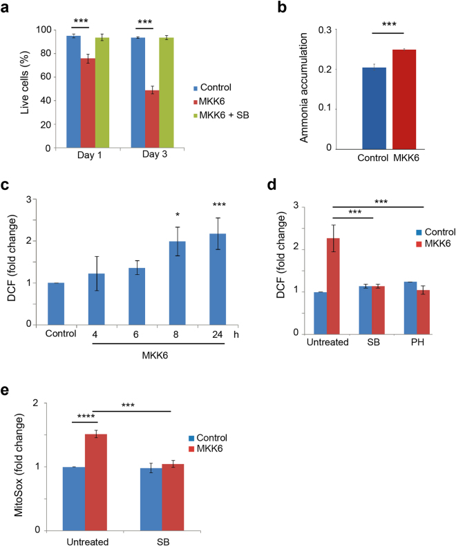
Figure 4.
Increased mitochondrial ROS contributes to cell death induced by MKK6 expression. U2OS cells expressing a Tet-regulated construct were either mock treated (control) or treated with tetracycline for the indicated times to induce the expression of constitutively active MKK6. (a) Cell survival was assayed at the indicated times using Annexin V/PI staining. Live cells were determined as the cell population Annexin V− and PI−. (b) Intracellular ammonia accumulation was measured in control and MKK6 expressing cells at 32 h and is presented as nmol / 5 × 10⁴ cells. (c) Total ROS levels were analysed at the indicated times after MKK6 induction using the DCFH-DA probe. Values are presented as fold change of mean fluorescence of DCF versus the control cells. (d) ROS levels in control and MKK6 expressing cells treated for 24 h with the p38α inhibitors PH797804 (PH) or SB203580 (SB) were analysed using the DCFH-DA probe. Values are presented as fold change in the mean fluorescence of DCF. (e) Mitochondrial ROS levels in control and MKK6 expressing cells either mock-treated or treated with SB were evaluated at 24 h using the MitoSox probe. Values are represented as fold change in the mean fluoresce of MitoSox normalized towards control cells.

