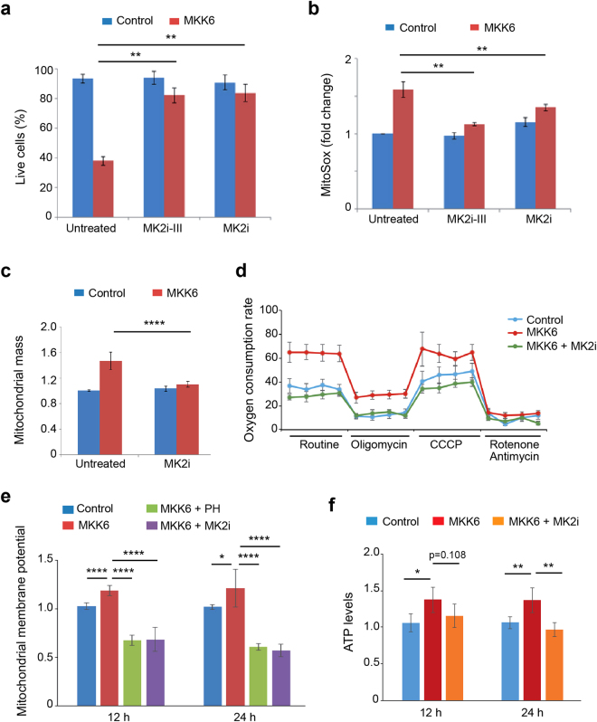
Figure 7.
MK2 mediates changes in mitochondrial function induced by MKK6 expression. U2OS cells expressing a Tet-regulated construct were either mock treated (control) or treated with tetracycline for the indicated times to induce the expression of constitutively active MKK6. (a) Control and MKK6 expressing cells were grown in complete medium in the presence or absence of the MK2 inhibitors PF3644022 (MK2i) or MK2 inhibitor III (MK2i-III) for 3 days, and cell survival was assayed using Annexin V/PI staining. Live cells were determined as the cell population Annexin V− and PI−. (b) Control and MKK6 expressing cells were incubated with MK2i or MK2i-III for 24 h, and mitochondrial ROS levels were analysed using the MitoSox probe. Values are represented as fold change in the mean fluorescence of MitoSox normalized towards control. (c) Control and MKK6 expressing cells were incubated with MK2i for 24 h and mitochondrial mass was analysed by Mitotracker Deep Red. Values are represented as fold change versus the control. (d) The oxygen consumption rate was analysed 24 h after the induction of MKK6 expression in the presence or absence of MK2i. Results are presented as pmoles of consumed O2 per 105 cells. (e) Mitochondrial membrane potential in control and MKK6 expressing cells in the presence or absence of PH or MK2i was measured at the indicated times. (f) ATP levels were analysed at the indicated times after MKK6 induction in the presence or absence of MK2i, and were normalized towards the cell number.

