Figure 2.
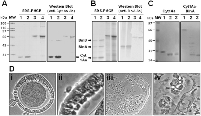
Protein profile and antigenicity of the Cyt1Aa-BinA chimera. Inclusion bodies were purified from Bacillus thuringiensis subsp. israelensis 4Q7 strains producing (A) Cyt1Aa (4Q7/pWF45; lanes 1, 2), Cyt1A-BinA (4Q7/pBU-cyt1Aa-binA; lanes 3, 4), (B) Cyt1Aa and BinAB (4Q7/45S1; lanes 1, 2) or Cyt1A-BinA (4Q7/pBU-cyt1A-binA; lanes 3, 4). Inclusions were solubilized and fractionated by SDS-PAGE in a 10% gel and electroblotted for Western analysis using rabbit anti-Cyt1Aa and anti-BinA antibodies (Ab). Lanes 1, 2, and 3, 4, respectively, 0.75 μg and 1.5 μg of protein; molecular masses: Cyt1Aa, 27.2 kDa; BinA, 42 kDa; BinB, 52 kDa; and Cyt1Aa-BinA 69.6 kDa. (C) SDS-PAGE demonstrating proteolytic cleavage of Cyt1Aa-BinA by trypsin, with Cyt1Aa as a control. Purified parasporal inclusions were solubilized in 50 mM NaOH, supernatants collected and neutralized with HCl, and digested with the enzyme at 28 °C. Untreated samples, 1.5 hr (lane 1), and trypsin-treated samples, 0.5 hr (lane 2) and 1.5 hr (lane 3). (D) Midgut histopathology caused by Cyt1Aa-BinA chimera in fourth instars of Culex quinquefasciatus 8 hours post-treatment at the LC95 concentration; Control midgut epithelium, (i) and (ii), respectively, 100x and 400x magnification. Midgut epithelium of a treated larva (iii) and (iv), respectively 100x and 600x magnification. Note the vacuoles in cells designated by arrows in D (iv) that have sloughed from the midgut basement membrane (C, 100x; D, 600x). The central circular area in A is the food column surrounded by the peritrophic membrane. MW, protein molecular mass standards; kDa, kilodaltons.

