Figure 4.
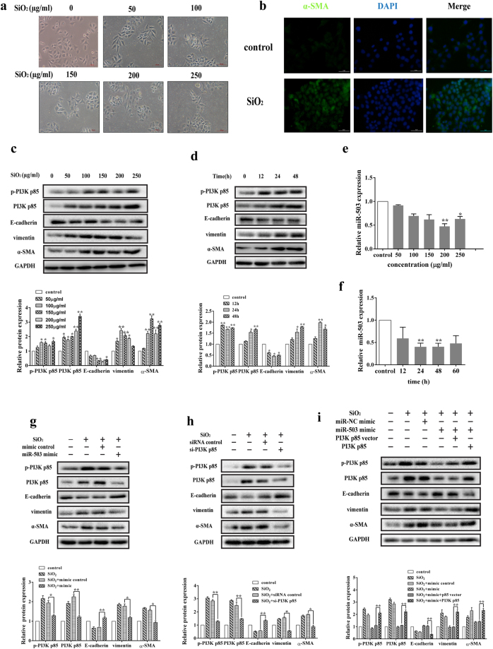
MiR-503 blocks the process of EMT via targeting PI3K p85 in HBE cells. (a) The cells (HBE) morphology shifted toward mesenchymal-like phenotype when treated with different concentrations of silica suspension (0, 50, 100, 150, 200, 250 μg/ml) for 48 h. The pictures were captured by the inverted microscope (Olympus). The scale bar is 100 μm. (b) The fluorescence intensity of α-SMA in HBE cells treated with 200 μg/ml silica was notably higher than the control group. The scale bar is 50 μm. (c,d) The protein expression levels of p-PI3K p85, PI3K p85, vimentin and α-SMA were obviously elevated and the E-cadherin protein levels were decreased when treated with different doses of silica for 48 h and 200 μg/ml silica for different time points. And the relative protein expression levels were analyzed by the ImageJ program. *P < 0.05 and **P < 0.01 versus the control group. (e,f) The miR-503 expression levels were significantly decreased in HBE cells treated with different doses of silica for 48 h and 200 μg/ml silica for different time points, *P < 0.05 and **P < 0.01 versus the control group. (g) MiR-503 mimics were transfected to HBE cells one day before treated with silica and then cultured for 48 h. MiR-503 mimics reversed the protein expression levels of the target PI3K p85 and EMT markers (E-cadherin, vimentin, α-SMA) in HBE cells, *P < 0.05 and **P < 0.01 versus the SiO2 + miR-NC mimic group. (h) The siRNA of PI3K p85 reduced the protein expression of PI3K p85 and alleviated the process of epithelial mesenchymal transformation, *P < 0.05 and **P < 0.01 versus the SiO2 + siRNA control group. (i) Overexpression of PI3K p85 significantly counteracted the inhibitory effects of miR-503 in the process of EMT by rescue experiment, **P < 0.01 versus the SiO2 + mimic + p85 vector group.

