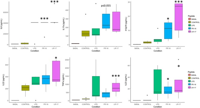
FIGURE 2.

Main cytokine levels affected by the presence of peptides LR17 and FR16. Boxplots represent median and interquartile range for the selected cytokines (see all in Supplementary Figure S1). These were quantified in the supernatants after 5 days of in vitro co-culture of human PBMCs and the bacterial peptides FR-16 and LR-17, from B. longum DJO10A and B. fragilis YCH46, respectively. Significant differences were assessed by the non-parametric Mann–Whitney U test and are represented with ∗, ∗∗, or ∗∗∗ (p < 0.05, 0.01, or 0.001 respectively). Significant differences refer to control conditions (PBMCs + anti-CD3).
