Fig. 4.
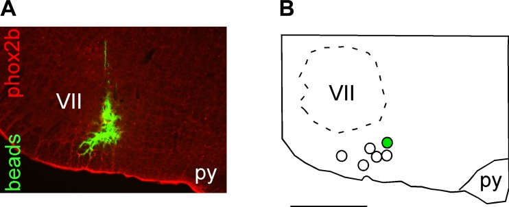
Histological confirmation of injection sites in the RTN. A: photomicrograph of a typical injection of fluorocitrate (FCt) into the RTN. Note that the FCt injection (demonstrated by presence of green fluorescent microbeads) targets the region that contains the highest density of phox2B immunoreactive neurons (in red). B: computer-assisted plots of the center of the FCt injection sites targeted to the RTN revealed by the presence of fluorescent microbeads. All sites are projected on a single section located at Bregma −11.6 mm according to Paxinos and Watson (1998). py, Pyramidal tract; VII, facial motor nucleus. Scale, 1 mm.

