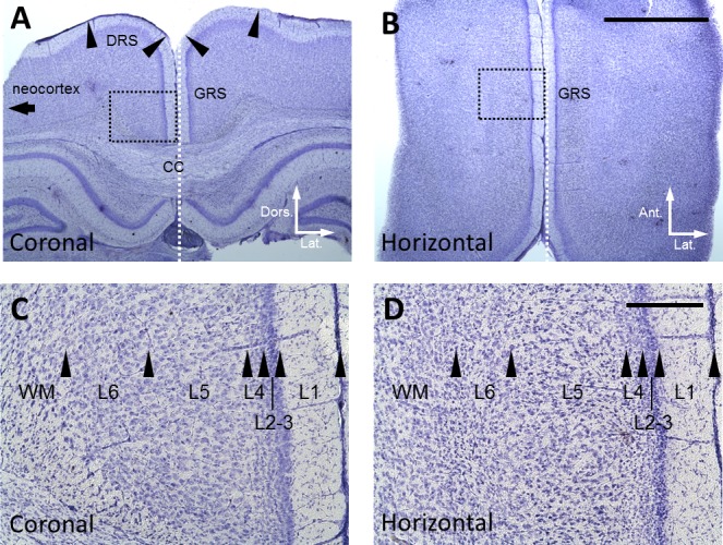
Fig. 1.

Location of recording sites. A: Nissl-stained coronal section of the GRS. DRS, dysgranular retrosplenial cortex; and CC, corpus callosum. Arrowheads indicate the borders of cortical areas. B: Nissl-stained horizontal section of the GRS. Scale bar, 2 mm, is also applied to A. White dotted line in A and B shows the midline of the brain. C and D: Magnified view of the area indicated by black dotted rectangle in A and B: respectively. Arrowheads indicate the borders of cortical layers. Scale bar, 200 µm, in D is also applied to C.
