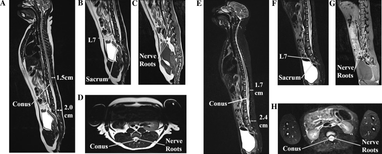
Fig. 1.
MRI of a female (A–D) and male (E–H) rhesus macaque. A and E: sagittal T2 sequences of the entire spinal column. The conus medullaris is seen at L3–L4, and the depth of the lumbar epidural space ranges from 1.5 to 2.4 cm. B and F: sagittal lumbar T2 sequences showing the transition from lumbar to sacral vertebrae. C and G: parasagittal (just off midline) lumbar T1 sequences showing the nerve roots exiting out their respective neural foraminae. D and H: axial lumbar T2 sequences showing the conus medullaris and surrounding nerve roots (which become the cauda equina inferiorly).

