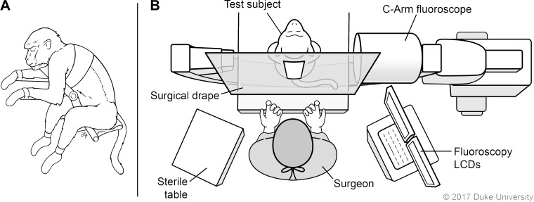
Fig. 2.
Surgical setup for implantation of epidural catheter-port. A: the monkey is positioned upright and with back in flexion. B: the c-arm is placed under the sterile drape so images can be acquired during placement. LCD, liquid crystal display computer monitor. Illustrated by Lauren Halligan, MSMI; copyright Duke University; with permission under a CC BY-ND 4.0 license.

