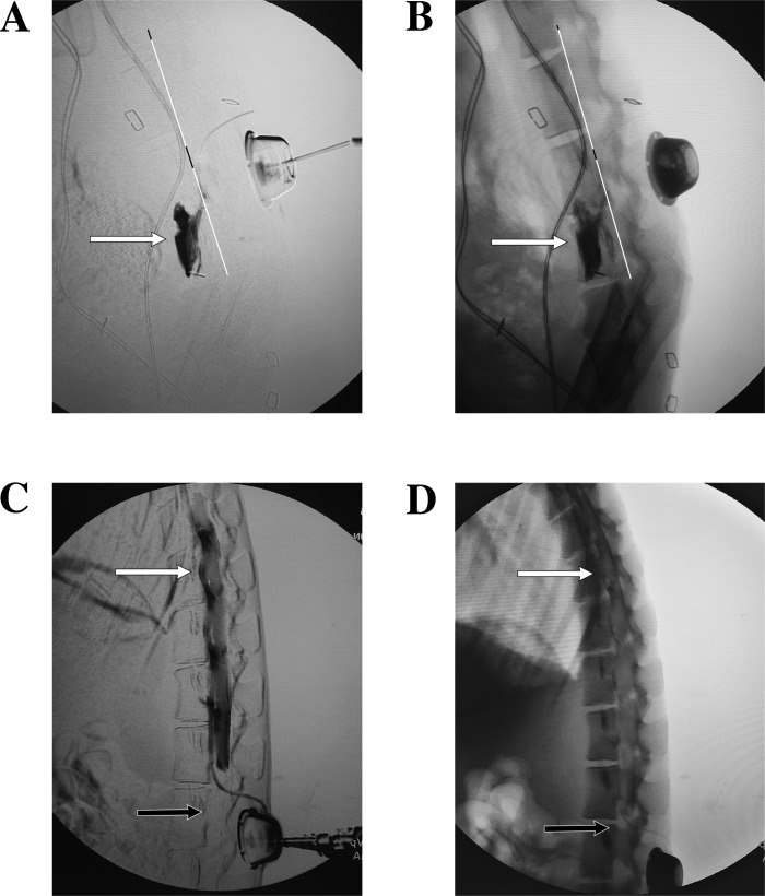
Fig. 8.
Aberrant dye distributions. A and B: subtracted (A) and nonsubtracted (B) fluoroscopic X-rays showing dye leaving the L5–L6 neural foramen. Note the dye is collecting anterior to the dorsal aspect of the vertebral body (white line) and not in the epidural space, suggesting the catheter has followed the nerve root out that foramen. C and D: subtracted (C) and nonsubtracted (D) fluoroscopic X-rays showing the catheter in the ventral epidural space at L3; however, dye is noted to be spreading far more cephalad (white arrows) than caudally (black arrows). Compare these results with Fig. 3, H and I.

