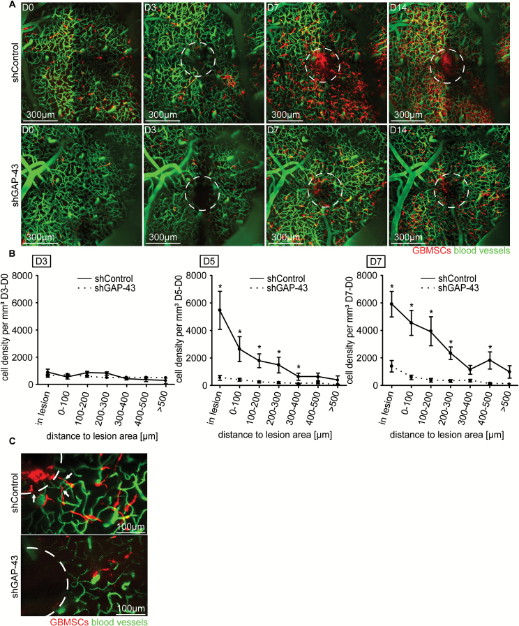
Fig. 3.
GAP-43 deficient glioma cells fail to repopulate surgical lesions. (A) Representative in vivo MPLSM 3D images of the repopulation of lesioned areas (dotted circles) by S24 shControl vs shGAP-43 GBMSCs, up to 14 days after surgical lesion, 75–225 µm depth under the brain surface. (B) Quantification of the tumor cell number in S24 shControl GBMSCs vs S24 shGAP-43 cells over time (9–1467 GBMSCs quantified; 4 regions, n = 3 mice per group, t-test). (C) Representative in vivo MPLSM image of S24 tumor cells repopulating the lesioned area (dotted circle) in shControl vs shGAP-43 on day 5 after lesion. Note the TMs reaching toward the lesion in shControl and their absence in shGAP-43. Error bars show SEM. *P < 0.05.

