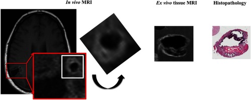
Fig. 6.
Correlation among in vivo MRI-ex vivo MRI-histopathologic correlation targeting on the tissue A with heavy calcified nodular formation (rectangle in white). In the magnified view of the area with red rectangle, a nodule with MR signal characteristics for heavy calcification is demonstrated, which is most compatible with the tissue A in ex vivo MRI and histopathology. On in vivo MRI, the area highlighted in a white square is cropped and rotated in a position that provides a similar appearance with ex vivo MRI and histopathology. Correlation for the tissue B is difficult due to fragmented nature of the neurosurgical specimen, respectively.

