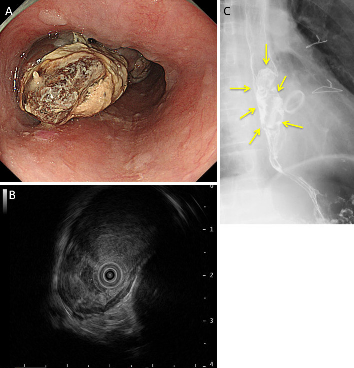
Figure 1.
Endoscopic image (A), endoscopic ultrasonography (B), and esophageal barium examination (C) of the patient. An elevated lesion with an lobulated-black tone in the mid-thoracic esophagus at a site 37-43 cm from the incisors was revealed, with some areas with a white moss appearance (A). Endoscopic ultrasonography demonstrated a low echoic legion, a solid tumor mass, in the esophageal wall (B). Esophageal barium examination showed rising, steep ridged tumors that were localized in the mid-esophagus of the chest (C).

