
Figure 1.
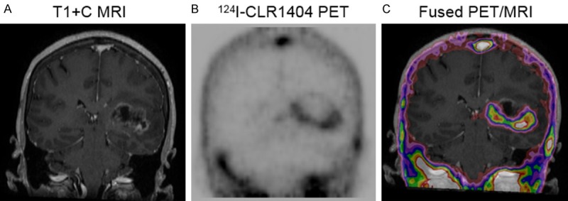
Patient with newly diagnosed high grade tumor (WHO Grade 4 glioma). A: T1 contrast-enhanced coronal MRI demonstrating heterogeneous tumor enhancement. B: Corresponding 124I-CLR1404 coronal PET with intense uptake laterally, inferiorly and medially, minimal uptake superiorly and no uptake centrally. C: Corresponding fused PET/MRI, with PET uptake extending slightly beyond MRI enhancement, except superiorly.