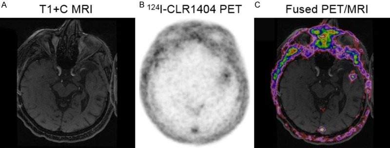
Figure 4.

Patient with recurrent low grade tumor (WHO Grade 2 glioma). A: Axial T1 contrast MRI demonstrates a prior resection cavity in the anterior left temporal lobe but no enhancement. B: 124I-CLR1404 axial PET demonstrates mild uptake adjacent to the posterior resection cavity without enhancement on MRI. C: Fused PET/MRI image further depicts these findings.
