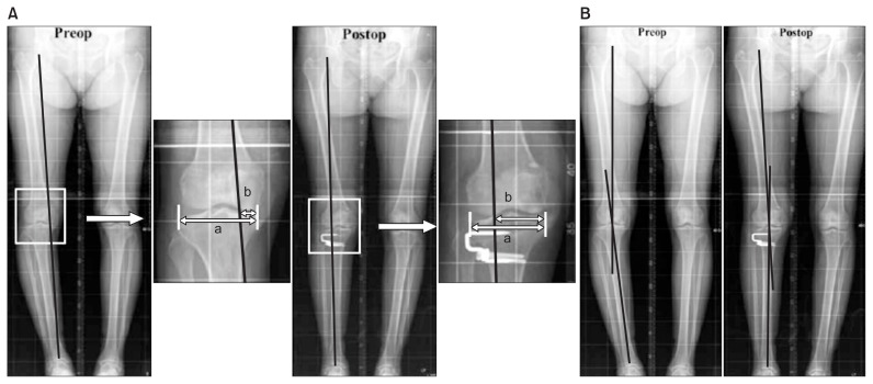
Fig. 1.
Radiographic measurement of preoperative (preop) and postoperative (postop) mechanical axis (MA) and the percentage of mechanical axis (MA%). (A) The MA% was evaluated on the orthoroentgenogram and expressed in percentage [(b/a)×100]. (B) The MA was defined as the angle between femoral and tibial mechanical axes on the orthoroentgenogram. a: the width of tibia plateau, b: the distance from the medial border of the medial tibial condyle to the point at which the mechanical axis intersects the knee joint line.

