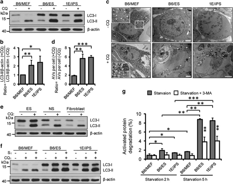
Figure 1.
High autophagic flux in pluripotent stem cells. (a) Western blotting for LC3 and β-actin in B6/MEF, B6/ES, and 1E/iPS cells. β-Actin served as the loading control. CQ, chloroquine. CQ treatment: 50 μM, 5 h. (b) Quantification of autophagic flux by calculating the ratio of LC3-II with or without CQ treatment in (a). Data shown are the mean±S.D., n=4, *P<0.05, **P<0.01 (Student’s t-test). (c) Representative TEM images of autophagic vacuoles (AVs, indicated by white arrowheads) in B6/MEF, B6/ES, and 1E/iPS cells with or without CQ treatment (50 μM, 5 h); scale bars, 2 μm. (d) Quantification of autophagic flux calculated from the ratio of the number of AVs in untreated and CQ treated cells in (c). Mean±S.D. from three independent experiments are shown and ~20 cells were counted. **P<0.01, ***P<0.001 (Student’s t-test). (e) Western blotting for LC3 and β-actin in ESC, ESC-derived NSC, and ESC-derived fibroblast. β-Actin served as the loading control. CQ, chloroquine. CQ treatment: 50 μM, 5 h. (f) Western blotting for LC3 and actin in B6/MEF, B6/ES, and 1E/iPS cells under basal or starvation conditions. S, starvation. Cells underwent EBSS starvation for 5 h (these conditions were used for similar experiments). CQ treatment: 100 μM, 5 h. (g) Time-course analysis of degradation of long-lived proteins in B6/MEF, B6/ES, and 1E/iPS cells under starvation conditions (EBSS starvation for 2 or 5 h). Inhibition of degradation of long-lived proteins by treatment with 10 mM 3-methyladenine (3-MA) for 2 or 5 h. Data shown are mean±S.D., n=3, *P<0.05, **P<0.01, ***P<0.001 (Student’s t-test). Images in (a, e, and f) are representative of three independent experiments

