Figure 1.
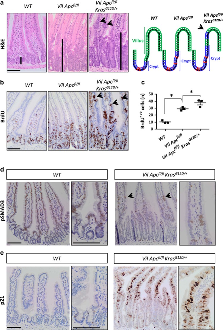
KrasG12D/+ enhances both the crypt-progenitor phenotype and transforming growth factor-β (TGFβ) pathway activation following Apc loss. (a) Haematoxylin and eosin (H&E) staining and a schematic model of the crypt-progenitor phenotype of wild-type (WT), VilCreERApcfl/fl (Vil Apcfl/fl) and VilCreERApcfl/flKrasG12D/+ (Vil Apcfl/flKrasG12D/+) small intestine. Note the presence of dedifferentiated cells (black arrow) in the villus of the Vil Apcfl/flKrasG12D/+genotype. Black bars highlight the differences in crypt length. (b) Bromodeoxyuridine (BrdU) staining of WT, Vil Apcfl/f and Vil Apcfl/flKrasG12D/+ small intestine. Note the presence of proliferating cells (black arrow) in the villi of the Vil Apcfl/flKrasG12D/+genotype. (c) Quantification of total BrdU+ cells demonstrating increased proliferation in the Vil Apcfl/flKrasG12D/+ genotype. Error bars represent mean± S.E.M., *P=0.04 Mann–Whitney test, one-tailed, n=3 biological replicates. (d) pSMAD3 and (e) p21 IHC analysis of intestine from WT and Vil Apcfl/flKrasG12D/+ mice. Black arrows indicate positive cells in the villi. High magnification insets highlight the presence of positive cells. Scale bars, 100 μm

