Figure 5.
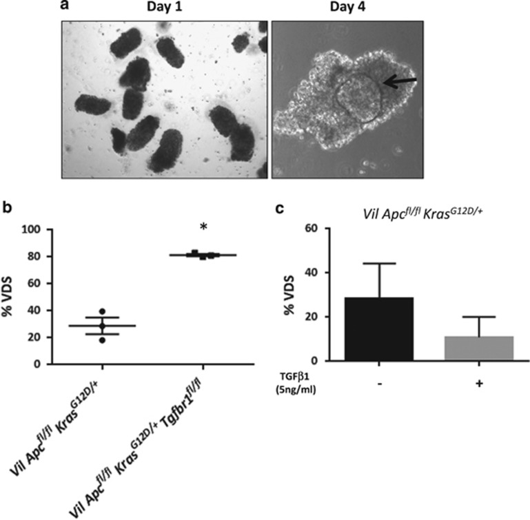
Transforming growth factor-β (TGFβ) signalling attenuates the capacity of villi to dedifferentiate in vitro. (a) Morphology of villi freshly purified from VilCreERApcfl/flKrasG12D/+ mice at day 1 (left panel) and day 4 (right panel) in culture. Black arrow indicates a spheroid inside the villus at day 4. (b) Quantification of VDS generated from Vil Apcfl/flKrasG12D/+ and VilCreERApcfl/flKrasG12D/+Tgfbr1fl/fl (Vil Apcfl/flKrasG12D/+Tgfbr1fl/fl) intestine. Error bars represent mean±S.E.M., *P=0.04 by Mann–Whitney test, one-tailed, n=3 biological replicates. (c) Quantification of VDS derived from VilCreERApcfl/flKrasG12D/+ villi treated with TGFβ1 (5 ng/ml) or Vehicle for 72 h. Plot represents mean with standard deviation (s.d.), n=3 biological replicates

