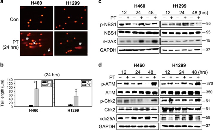
Figure 5.
PT-induced DNA damage in lung cancer cells. (a) Photomicrography of H460 and H1299 cells treated with 50 μM PT for 24 h and analyzed using comet assay. (b) DNA damage level expressed as tail length (μm) calculated using Komet 5.5 software (Kinetic Imaging Ltd., London, UK) after 24 h of 50 μM PT treatment in H460 and H1299 cells (mean±S.E.M., n=3, *P<0.05 compared with control groups). (c) H460 and H1299 cells were treated with 50 μM PT for 12, 24 and 48 h. Total-cell lysates were subjected to western blot analysis for (c) DNA damage proteins and (d) DNA sensor kinase (ATM), cell cycle checkpoint kinase (Chk2), and cell cycle regulatory protein (cdc25A). Membranes were probed with an anti-GAPDH antibody to confirm equal loading of proteins. Results are representative of three independent experiments

