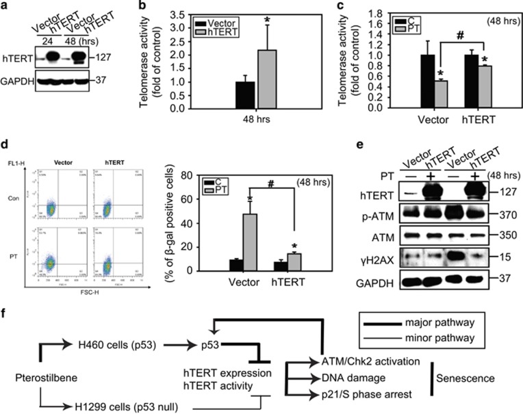
Figure 6.
Exogenous telomerase expression rescued telomerase activity and decreased senescence induced by PT. (a) H460 cells were transiently transfected with either pcDNA-3.1 vector or pcDNA-3.1 hTERT-3HA plasmid for 24 and 48 h. Cell lysates were analyzed for the expression of hTERT by western blot analysis and (b) telomerase enzyme activity. (c) The telomerase enzyme activity of the vector or hTERT transfected H460 cells treated with 50 μM PT for 24 h were measured using a TRAPeze RT Telomerase Detection Kit. (mean±S.E.M., n=3, *P<0.05, significantly lower than control groups. #P<0.05, significantly higher than vector groups). (d) The SA β-gal activity of cells treated as in (b) was stained with C12FDG and analyzed by flow cytometry. X axis: FSC-H, Y axis: FL1-H. Data represented the mean±S.E.M. of three independent experiments. *P<0.05, compared with control groups. #P<0.05, significantly lower than vector groups. (e) Cells were treated as described in (b) and immunoblotting was performed with anti-hTERT, p-ATM, ATM, γH2AX, and GAPDH antibodies. Results are representative of three independent experiments. (f) Proposed model summarizing PT-induced senescence in lung cancer cells. In p53 wild-type H460 cells, PT inhibits hTERT enzyme activity and protein expression resulting in the subsequent induction of DNA damage, activation of ATM/Chk2 and p53, and S phase arrest. Activation of p53 positive feedback provokes hTERT downregulation, resulting in senescence in H460 cells. Interestingly, PT slightly inhibited hTERT enzyme activity resulting in less senescence in H1299 cells, suggesting that PT-induced senescence in lung cancer cells partially through p53-mediated hTERT inhibition

