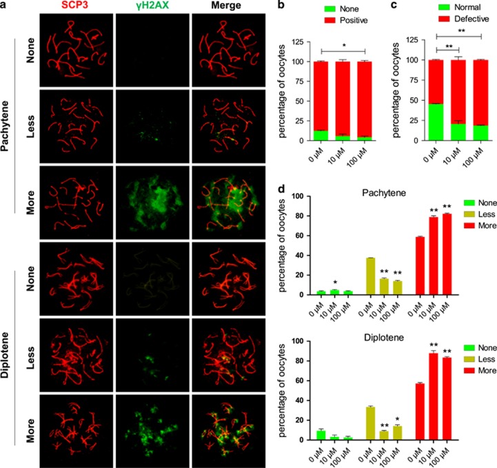
Figure 2.
Effect of DEHP on γH2AX pattern. (a) Immunolabeling of the oocyte chromosomes with anti-SYCP3 (red) and anti-γH2AX (green) antibodies. (b) Percentage of positive γH2AX oocytes (79.36±3.97% and 81.40±0.62%, respectively), were significantly higher than that of control group (54.70±0.61% P<0.01). (c) Percentage of normal, negative or weak γH2AX staining and defective (strong γH2AX staining) oocytes after six days of culture with or without DEHP. (d) Percentage of oocytes at the pachytene and diplotene stages showing negative, weakly and strongly positive γH2AX staining after six days of culture with or without DEHP. (*P<0.05; **P<0.01)

