Figure 7.
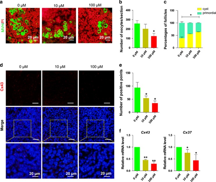
DEHP exposure causes a reduction of oocyte number and of cyst breakdown (a) Control and DEHP exposed ovarian tissue stained for MVH (green specific for oocytes) after 10 days DEHP exposure, nuclei red; bar is 20 μM. (b) Number of oocytes in control and DEHP exposed groups. (c) Percentage of oocytes in cysts and primordial follicles in control and DEHP exposed groups. DEHP impairs the expression of connexins, (d) Control and DEHP exposed ovarian tissues stained for Cx43 (red), nuclei blue; bar is 20 μM. (e) Number of Cx43 positive gap junction plaques in control and DEHP exposed ovarian tissues. (f) qRT-PCR for Cx43 and Cx37. Relative fold changes are presented as mean±SD. All experiments were repeated at least three times. (* P<0.05; ** P<0.01)

