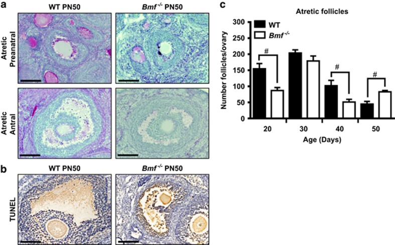
Figure 2.
Follicular atresia in ovaries from WT and Bmf−/− female mice. The numbers of atretic follicles were determined in ovaries from WT and Bmf−/− mice at PN20, 30 40 and 50 (n=6/age/genotype). (a) Representative images of atretic preantral (secondary) and antral follicles in PAS-stained ovarian sections from WT and Bmf−/− mice at PN50. Atretic preantral and antral follicles were characterised by the presence of degrading oocytes and/pyknotic granulosa cells. Scale bars=50 μm. (b) Representative images of TUNEL-positive atretic follicles (brown staining) in secondary follicles in ovarian sections from WT and Bmf−/− mice at PN50. Scale bars=50 μm. (c) Data are expressed as mean±S.E.M. #P<0.05 for comparison of Bmf−/− versus WT females at each age P<0.05 (two-tailed unpaired student’s t-test). For clarity, only pairwise comparisons between WT and Bmf−/− females at each age are shown. See Supplementary Tables 1 and 2 for statistical significance of all pairwise comparisons within a genotype

