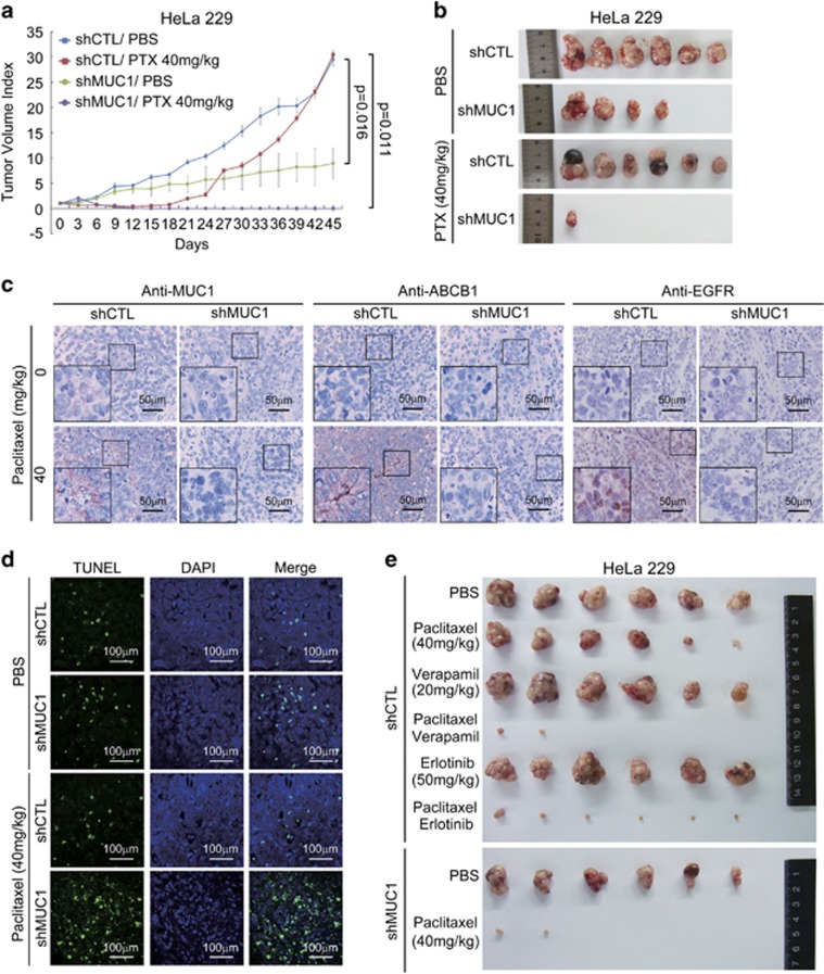
Figure 7.
Co-administration of the inhibitors of MUC1–EGFR–ABCB1 axis and PTX prevents tumor relapse. (a and b) Six-week-old female BALB/c nude mice were subcutaneously injected with 2.5 × 10⁶ HeLa229/shCTL cells or HeLa229/shMUC1 cells in ventral flanks. When tumor reached approximately 4 mm × 4 mm, the mice were injected intraperitoneally with PTX at 40 mg/kg every three days for 15 days. The tumor sizes were measured every 3 days following PTX treatment. The tumor volume was calculated according to the formula: V=length × width2/2. The data indicated mean with S.E.M. of six mice in each group. (b) At the 45th day, all mice were killed and tumors were excised and photographed. (c and d) IHC stainings (c) or TUNEL assay (d) of tumor tissue sections were carried out. (e) 2.5 × 10⁶ HeLa229/shCTL cells or HeLa229/shMUC1 cells were subcutaneously injected in ventral flanks of 6-week-old female BALB/c nude mice. When the tumor reached 4 mm × 4 mm, the mice were blindly allocated into six groups and injected with PTX (40 mg/kg) in combination with verapamil (20 mg/kg) or erlotinib (50 mg/kg) intraperitoneally every 3 days for 15 days. At the 36th day, all mice were killed and tumors were excised and photographed

