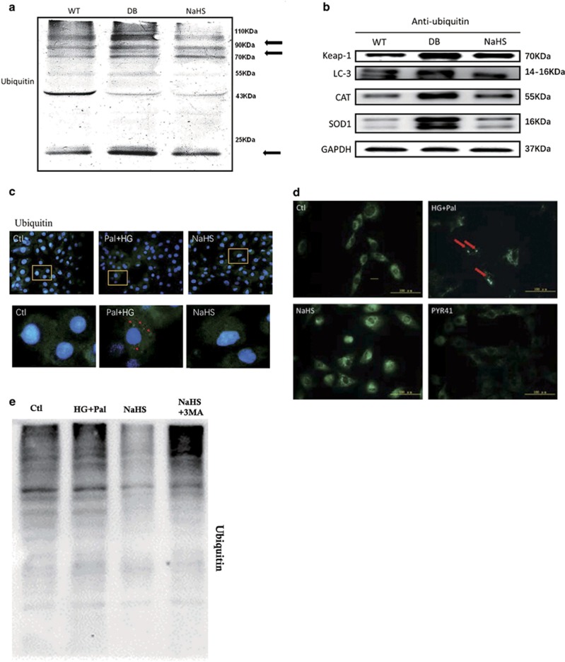
Figure 4.
Exogenous H2S attenuated ubiquitylation levels. (a) The expression of ubiquitous protein was analysed by western blotting in mouse myocardia. (b) The ubiquitylation level of Keap-1, SOD and CAT in mice myocardium was detected by immunoprecipitation and western blotting. H9C2 cells were treated with HG (40 mM)+palmitate (Pal, 200 μM), HG+Pal+NaHS (100 μM), HG+Pal+NaHS+3MA (2 mM, an inhibitor of autophagy) and HG+Pal+PYR41 (3 μM, an inhibitor of ubiquitin-activating enzyme (E1) for 48 h. (c) The ubiquitinated proteins in H9C2 cells were detected by an immunofluorescent assay (green); red arrows indicate ubiquitinated protein aggregates; scale bar: 100 μm. (d) The autophagosomes were detected by MDC test in H9C2 cells (green). Red arrows indicate autophagosome accumulation; scale bar: 100 μm. (e) The ubiquitinated protein level was detected by western blotting

