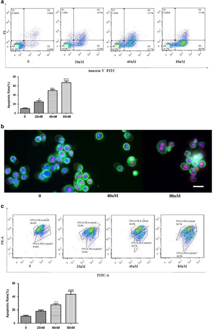
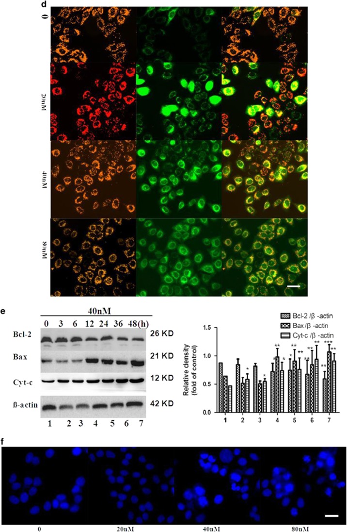
Figure 3.
Compound induced cell apoptosis. (a) SMMC-7721 cells treated with the title compound (20, 40, and 80 nm) for 48 h were stained with Annexin V/PI and then were analyzed using fluorescence cytometry and (b) fluorescence microscopy (magnification × 200). (c) Mitochondrial membrane depolarization analysis was performed by detecting the mitochondrial transmembrane potential using JC-1 stain treated with the compound for 48 h and then it was analyzed with fluorescence cytometry and (d) fluorescence microscopy (magnification × 200). (e) Western blot and densitometric analyses for Bcl-2 /Bax and cyt-c after SMMC-7721 cells treated with the title compound. (f) Compound induced nuclear chromatin condensation. SMMC-7721 cells were treated with 0, 20, 40, and 80 nm of the title compound for 48 h and then were stained with DAPI as described in Materials and Methods. Cells were examined and photographed using a fluorescence microscope at × 200. Representative blots were from three independent experiments. For the statistics of each panel in this figure, data are expressed as mean±S.D. (n=3); scale bars are 50 μm; *P<0.05 versus control, **P<0.01 versus control, ***P <0.001 versus control


