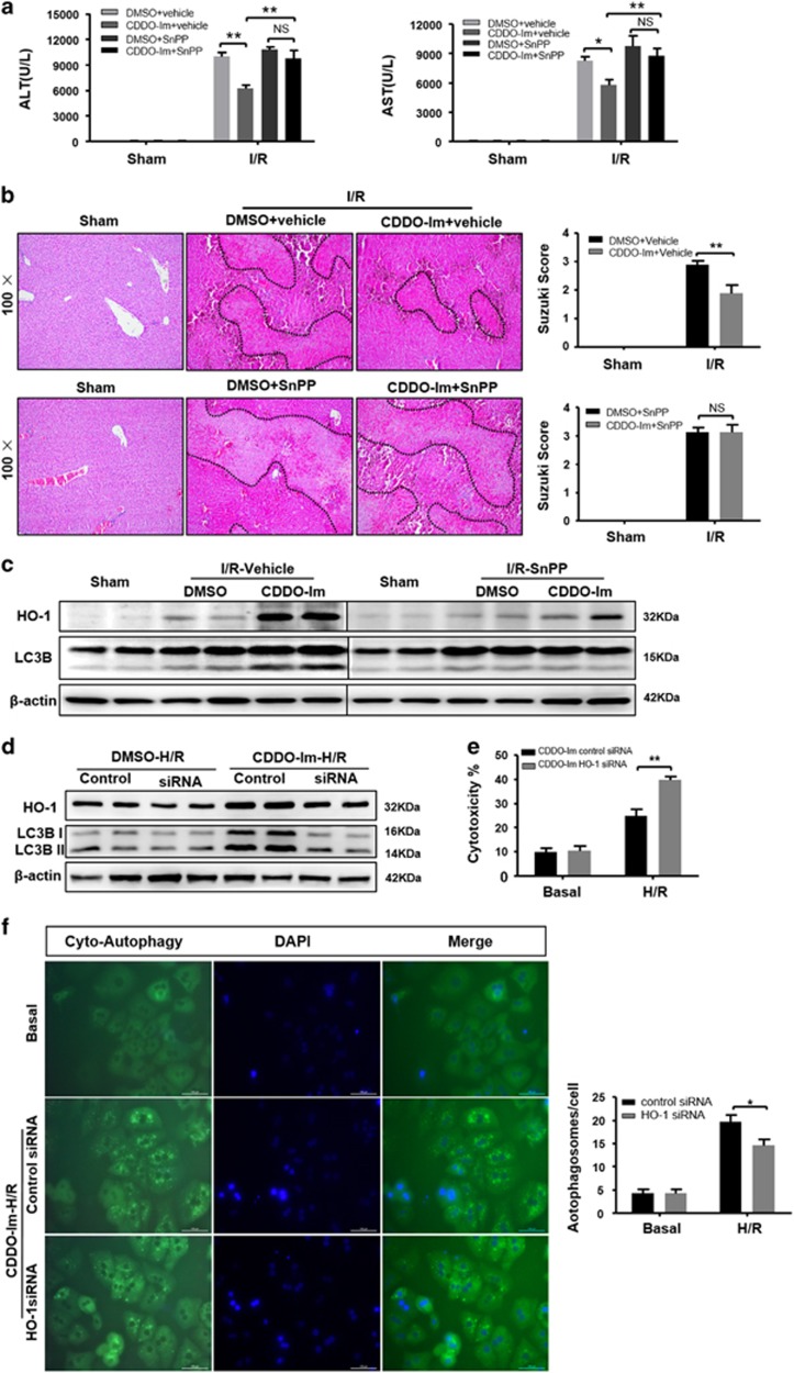
Figure 8.
CDDO-Im in induction of autophagy depends on induction of HO-1. (a–c) WT mice were pretreated with SnPP (50 mg/kg, IP) 1 h before CDDO-Im (2 mg/kg, IP) or DMSO and killed at 6 h after reperfusion. (a) Serum ALT and AST levels. (b) Representative hematoxylin and eosin (HE) stained sections (original magnification, × 100) and relevant average Suzuki score. (c) Western blot analysis of HO-1 and LC3B both in the SnPP and vehicle pretreatment with or without CDDO-Im mice livers at 6 h after reperfusion. (n=3-4 per group, mean±S.E.M., **P<0.01, *P<0.05). (d) Immunoblots indicating expression of HO-1 and LC3B protein after treatment with HO-1 siRNA in the presence and absence of CDDO-Im (200 μM) and densitometric analysis of LC3B-II and HO-1 expression. (e) Quantification of average cytotoxicity (% cell death) in different group were plotted. (f) Representative fluorescence micrographs display autophagosomes in primary hepatocytes with CDDO-Im in the presence HO-1 siRNA or control siRNA (original magnification, × 400). The numbers of autophagosomes were determined. (n=3-4 per group, mean±S.E.M., **P<0.01, *P<0.05)

