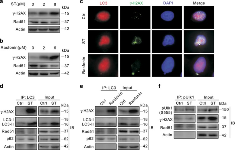
Figure 4.
LC3 interacts with γ-H2AX and Rad51. (a and b) 786-O cells were treated with ST (0–8 μM) or rasfonin (0–6 μM) for 3 h, cells were lysed and subjected to immunoblotting with the antibodies indicated. (c) 786-O cells were treated with ST or rasfonin for 3 h, and the images were obtained using fluorescence microscopy following staining with the antibodies of LC3 and γ-H2AX. (d–f) 786-O cells were incubated with ST or rasfonin for 3 h and lysed, and LC3s or pULK1s were precipitated using the antibody against LC3 or pULK1 (Ser555). The immunoprecipitates were resolved by electrophoresis and probed by immunoblotting with the indicated antibodies. All data were acquired from three independent experiments

