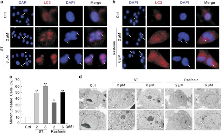
Figure 5.
ST and rasfonin increase the formation of micronuclei. (a and b) 786-O cells were treated with ST (0–8 μM) or rasfonin (0–6 μM) for 3 h, and the images were obtained by fluorescence microscopy after labeling the antibody of LC3 and DAPI with both 400 and 1000 magnification. The percentages of micronuclei were analyzed and shown in (c), and the data were presented as mean±S.D. in graphs, **P<0.01 versus control. (d) Electron microscopy was performed for 786-O cells following treatment with ST or rasfonin for 3 h. Representative images were presented, the arrows indicated micronuclei, and arrowheads showed the LC3 localized in micronuclei. Similarly experiments were carried out for three times

