Figure 1.
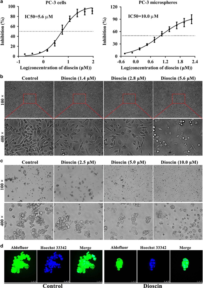
Dioscin exhibited cytotoxicity in PC3 cells, and disrupted mammospheres formation. (a) Effects of dioscin on the viabilities of PC3 cells and PC3 cell-derived mammospheres. (b) Effects of different concentrations of dioscin (1.4, 2.8 and 5.6 μM) for 24 h on the morphology and structure of PC3 cells (bright-field image). (c) Effects of different concentrations of dioscin (2.5, 5.0 and 10.0 μM) for 24 h on the morphology and structure of PC3 cell-derived mammospheres (bright-field image). (d) Mammosphere revealing cells staining positive for Aldefluor (green), counterstained with Hoechst 33342 (blue). Data are presented as the mean±S.D. (n=6)

